Osteoid Osteoma
A small and noncancerous bone tumor where the cells in the tumor, called osteoblasts, make new bone. There is no risk of the disease spreading.
A small and noncancerous bone tumor where the cells in the tumor, called osteoblasts, make new bone. There is no risk of the disease spreading.


A small and noncancerous bone tumor where the cells in the tumor, called osteoblasts, make new bone. It is almost always very painful and the pain is usually turned off by taking aspirin or NSAIDS. They are very irritating to the bone and result in significant swelling in the bone and surrounding tissues.


As the tumor slowly grows, the bone is weakened and you are at an increased risk of breaking the bone due to the tumor (called a pathological fracture). They may also spread to your lungs or other bones.
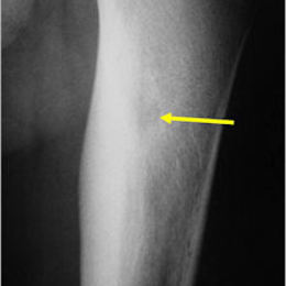
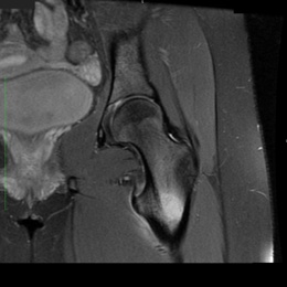
Radiographic imaging is used to help form a diagnosis. These include X-Ray, MRI, CT and Bone Scans.
An example of an osteoid osteoma is shown.
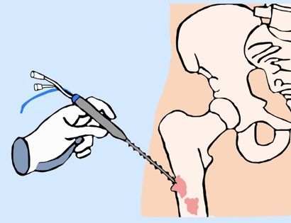
Treatment is usually with minimally invasive percutaneous CT guided radiofrequency ablation for osteoid osteomas in the long bones, pelvis and sometimes the spine. If not causing any symptoms, it may be recommended that the tumor be left alone.

The hands, wrist and feet and some other areas may be treated with an open surgical procedure called a “Burr-Down” resection where a high speed drill similar to a dental drill is used to shave the bone and remove the tumor.
I've seen many doctors and I can confidently attest Dr. Wittig is the preeminent orthopaedic specialist. He is genuinely kind and caring, as he demonstrated by completely addressing my concerns and compassionately relating to what I was dealing with. He clearly outlined the plan of attack, and recommended the two additional doctors who would become part of my 'team'. Dr. Wittig was so effective in allaying our fears and bringing us optimism. My surgery was significant, but I was up and walking the next day and back at the gym 5 weeks later. This is further testament to Dr. Wittig's skill. He saved my leg and my life, and I feel so very blessed to say he is my doctor. I have already recommended him to others, and I will continue to do so. I would trust him with my closest family and lifelong friends. BEST DOCTOR EVER.
S.G.
Myself and my amazing team are dedicated to saving your life and your limb. Losing a limb because of a tumor can be a terrifying experience. But, it does not have to be the only option. I’ve spent 20+ years as a Board-Certified Orthopedic Surgeon and Orthopedic Oncologist.
I’ve devoted my career to helping children and adults afflicted with bone and soft tissue masses by performing complex limb saving surgeries. Most patients can have their limb saved, which may require innovative techniques.
Patients afflicted with musculoskeletal tumors have complex conditions that are best taken care of at large hospitals. I am the Chairman of Orthopedics and Chief of Orthopedic Oncology at Morristown Medical Center. My philosophy is a multidisciplinary team approach, working together to tailor treatment to individual patients. Education and research are essential to my practice, providing the best setting for extraordinary patient care. Because of this, we have some of the top results in the country.