Chondromyxoid Fibroma
A Chondromyxoid Fibroma (CMF) is a slow-growing benign yet aggressive bone tumor or neoplasm. Surgery is the standard of treatment. It is important that it is removed because it can be spread to other body parts.
A Chondromyxoid Fibroma (CMF) is a slow-growing benign yet aggressive bone tumor or neoplasm. Surgery is the standard of treatment. It is important that it is removed because it can be spread to other body parts.


This is a benign aggressive, extremely rare bone tumor that is made up of three types of tissue: cartilage tissue, fibrous tissue and myxoid tissue. Aggressive means this tumor spreads and destroys bone. Benign means does not spread to other parts of the body. Myxoid tissue is a type of tissue that produces a mucous like substance. Fibrous tissue is made of collagen and forms tendons and ligaments as well as other soft tissues in the body. This is a slow-growing tumor that occurs primarily in the shin bone (tibia), femur, pelvis and small bones of hands and feet. This tumor grows and destroys bone but does not spread to other body parts. Surgery is the mainstay of treatment patients do not require any sort of chemotherapy or radiation.

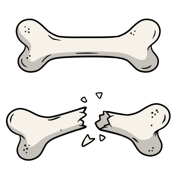
CMFs are benign tumors that will grow slowly over time. As the tumor grows, the bone is weakened and you are at an increased risk of breaking the bone due to the tumor (called a pathological fracture).
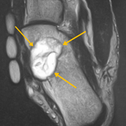
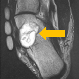
Radiographic imaging is used to help form a diagnosis of CMF. These include, X-Ray, MRI, CT and Bone Scans. An example of a CMF MRI is shown.
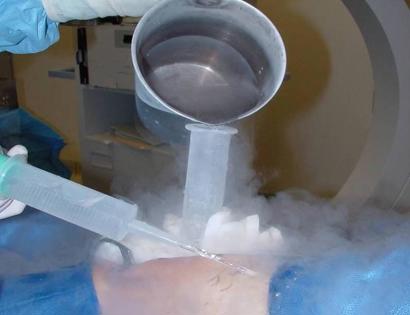
The primary form of treatment for CMF is surgical removal (excision). Most CMFs can be treated with a procedure called intralesional curettage and burr drilling called a curettage-resection.
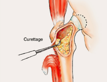
The primary form of treatment for CMF is surgical removal (excision). Most CMFs can be treated with a procedure called intralesional curettage and burr drilling called a curettage-resection. Curettage means to scoop the tumor out using a spoon-like tool called a curette. The burr-drilling is similar to a dental drill and removes additional tumor cells by shaving the bone in the cavity even further.
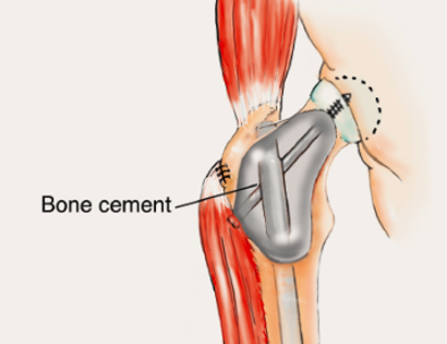
The empty bone cavity is usually filled with bone graft or bone cement. Bone can be donated (allograft) or taken from the patient themselves (autograft). Fixation devices, such as a plate and screws, may be used in specific situations to prevent postoperative fracture.
I've seen many doctors and I can confidently attest Dr. Wittig is the preeminent orthopaedic specialist. He is genuinely kind and caring, as he demonstrated by completely addressing my concerns and compassionately relating to what I was dealing with. He clearly outlined the plan of attack, and recommended the two additional doctors who would become part of my 'team'. Dr. Wittig was so effective in allaying our fears and bringing us optimism. My surgery was significant, but I was up and walking the next day and back at the gym 5 weeks later. This is further testament to Dr. Wittig's skill. He saved my leg and my life, and I feel so very blessed to say he is my doctor. I have already recommended him to others, and I will continue to do so. I would trust him with my closest family and lifelong friends. BEST DOCTOR EVER.
S.G.
Myself and my amazing team are dedicated to saving your life and your limb. Losing a limb because of a tumor can be a terrifying experience. But, it does not have to be the only option. I’ve spent 20+ years as a Board-Certified Orthopedic Surgeon and Orthopedic Oncologist.
I’ve devoted my career to helping children and adults afflicted with bone and soft tissue masses by performing complex limb saving surgeries. Most patients can have their limb saved, which may require innovative techniques.
Patients afflicted with musculoskeletal tumors have complex conditions that are best taken care of at large hospitals. I am the Chairman of Orthopedics and Chief of Orthopedic Oncology at Morristown Medical Center. My philosophy is a multidisciplinary team approach, working together to tailor treatment to individual patients. Education and research are essential to my practice, providing the best setting for extraordinary patient care. Because of this, we have some of the top results in the country.