Chondroblastoma
Chondroblastomas are a rare type of noncancerous (benign) bone tumor that begins in is made up of cartilage cells. Chondroblastomas grow at the ends of the body’s long bones (called the epiphysis) near the joints.
Chondroblastomas are a rare type of noncancerous (benign) bone tumor that begins in is made up of cartilage cells. Chondroblastomas grow at the ends of the body’s long bones (called the epiphysis) near the joints.


A rare type of noncancerous (benign) bone tumor that begins in is made up of primitive (The type of cells found in an embryo called chondroblasts) cartilage cells. The tumor is benign but also painful, aggressive and destroy bone and sometimes the adjacent joint as it grows. Chondroblastomas grow at the ends of the body’s long bones (called the epiphysis) near the joints. Common bones include the femur, tibia and humerus. They sometimes go undiagnosed and are attributed to sports injuries, growing pains and since adjacent to a joint, forms of arthritis.


If chondroblastomas are left untreated, they can continue to grow and destroy the surrounding bone, causing pain and sometimes a fracture.
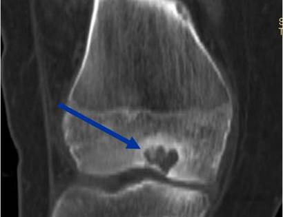
Radiographic imaging is used to help form a diagnosis of Chondroblastoma. Scans include X-Ray, MRI and Bone Scan. CT scan is sometimes used to assess the adjacent bone quality and look for flecks of calcium in the tumor to aid in diagnosis. MRI usually shows extensive swelling around the tumor that is sometimes misinterpreted as a cancer.
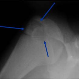
An example of a Chondrosarcoma X-Ray.
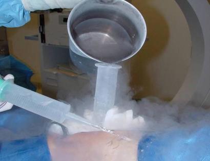
Treatment most commonly involves surgery to remove the tumor and to prevent damage to the nearby joint. Treatment usually involves curettage, midas rex burring and bone graft. Cryosurgery is often used by Dr. Wittig to kill microscopic cells and reduce the chances of the tumor coming back. Bone cement may also be used to fill the hole in certain instances. Radiofrequency ablation has also been described but is rarely used. Resection may be used when extremely extensive or recurrent. This includes removing an entire section of the bone.
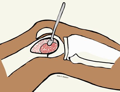
Intralesional Curettage means to scoop the tumor out using a spoon-like tool called a curette. This is a surgery that aims to remove the mass and restore the bone so that the patient can get back to normal function. The chondroblastoma is identified within the bone and scooped, or curetted, out. The cavity is then shaved down with a Midas Rex Drill, which is similar to a dental drill. This drill removes more tumor cells.
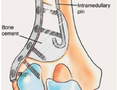
The empty bone cavity is usually filled with bone graft or bone cement. Bone can be donated (allograft) or taken from the patient themselves (autograft). Fixation devices, such as a plate and screws, may be used in specific situations to prevent postoperative fracture.
I've seen many doctors and I can confidently attest Dr. Wittig is the preeminent orthopaedic specialist. He is genuinely kind and caring, as he demonstrated by completely addressing my concerns and compassionately relating to what I was dealing with. He clearly outlined the plan of attack, and recommended the two additional doctors who would become part of my 'team'. Dr. Wittig was so effective in allaying our fears and bringing us optimism. My surgery was significant, but I was up and walking the next day and back at the gym 5 weeks later. This is further testament to Dr. Wittig's skill. He saved my leg and my life, and I feel so very blessed to say he is my doctor. I have already recommended him to others, and I will continue to do so. I would trust him with my closest family and lifelong friends. BEST DOCTOR EVER.
S.G.
Myself and my amazing team are dedicated to saving your life and your limb. Losing a limb because of a tumor can be a terrifying experience. But, it does not have to be the only option. I’ve spent 20+ years as a Board-Certified Orthopedic Surgeon and Orthopedic Oncologist.
I’ve devoted my career to helping children and adults afflicted with bone and soft tissue masses by performing complex limb saving surgeries. Most patients can have their limb saved, which may require innovative techniques.
Patients afflicted with musculoskeletal tumors have complex conditions that are best taken care of at large hospitals. I am the Chairman of Orthopedics and Chief of Orthopedic Oncology at Morristown Medical Center. My philosophy is a multidisciplinary team approach, working together to tailor treatment to individual patients. Education and research are essential to my practice, providing the best setting for extraordinary patient care. Because of this, we have some of the top results in the country.